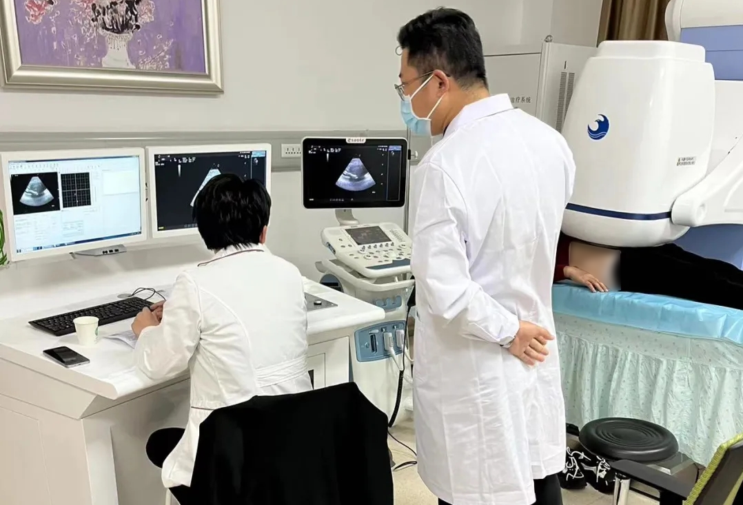
Clinical Cases for HY2900 HIFU
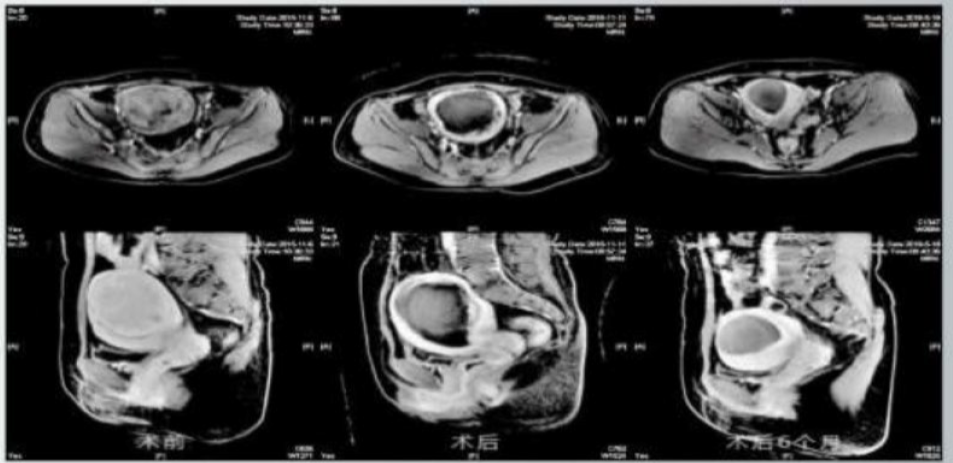
Patient 1 —— single posterior wall fibroid
Progressive volume reduction of a single posterior wall fibroid with sustained non-perfusion at 6-month follow-up, consistent with effective coagulative necrosis and tissue resorption.
Age: 47
Diagnosis: Uterine fibroid
Symptoms: Heavy menstrual flow, shortened menstrual cycle, prolonged menstrual period, anemia

POST-TREATMENT
6-MONTH FOLLOWUP
BASELINE
MRI Comparison:
BASELINE Lesion in the posterior uterine wall, approximately 78×75×68 mm, well‑defined low‑echo area with inhomogeneous moderate contrast enhancement.
POST-TREATMENT Posterior uterine wall lesion approximately 78×76×70 mm with a well‑defined, round non‑perfused region of 76×75×70 mm and moderate peripheral enhancement on contrast scan.
6-MONTH FOLLOWUP Posterior uterine wall lesion reduced to approximately 38×38×35 mm, with a well‑defined, round non‑perfused region of 38×38×32 mm and moderate peripheral enhancement on contrast scan.
Volume reduction from 78×75×68mm to 38×38×35mm.
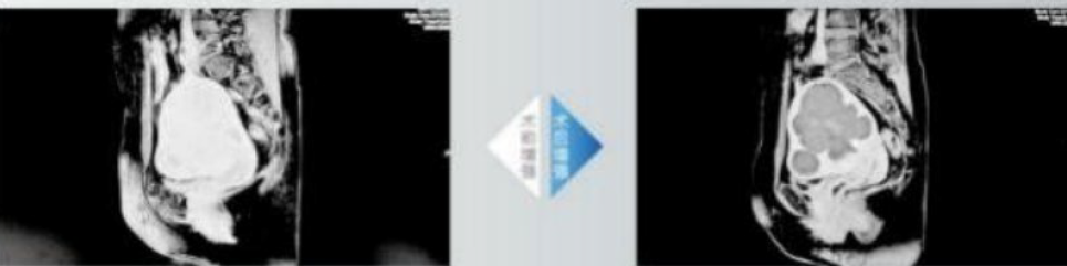
Patient 2 —— multiple fibroids
Successful treatment of multiple fibroids with immediate absence of contrast enhancement in treated lesions, indicating complete ablation despite complex fibroid distribution.
BASELINE
Age: 49
Diagnosis: Multiple uterine fibroids
Symptoms: Prolonged menstrual periods with heavy flow, moderate anemia.
POST-TREATMENT
MRI Comparison:
BASELINE Distorted uterine contour with multiple round abnormal signals of varying sizes in the intramural and subserosal layers, largest about 50×47 mm, moderately well‑defined with moderate progressive contrast enhancement.
POST-TREATMENT After contrast administration, no obvious enhancement in the lesions; the largest lesion (about 54×51 mm) shows clear coagulative necrosis compared with pre‑treatment.
Absent enhancement confirms clear coagulative necrosis of the largest lesion.
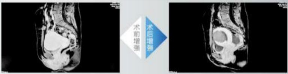
Patient 3 —— adenomyosis
Treatment of adenomyosis with significant reduction in lesion enhancement, correlating with expected symptom relief in this chronic condition.
Age: 41
Diagnosis: Uterine adenomyoma
Symptoms: Dysmenorrhea for over 3 years, heavy menstrual flow, moderate anemia.
BASELINE
POST-TREATMENT
MRI Comparison:
BASELINE Lesion in the anterior uterine wall approximately 66×51×43 mm, round abnormal signal, poorly defined, with marked enhancement after contrast administration.
POST-TREATMENT Minimal enhancement within the lesion after contrast-enhanced scan, size approximately 61×49×40 mm.
Lesion shrinkage from 66×51×43 mm to 61×49×40 mm indicates effective ablation and treatment response.
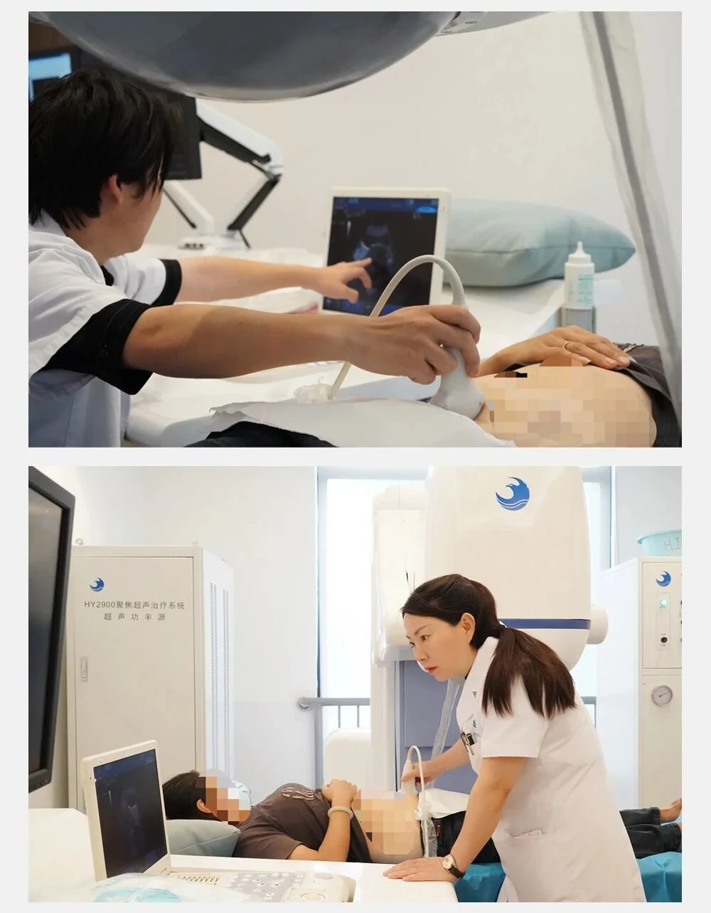
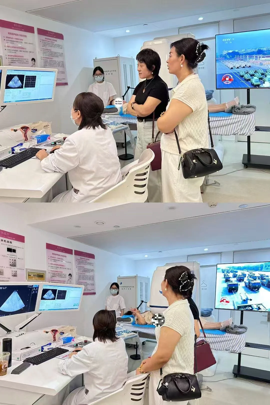
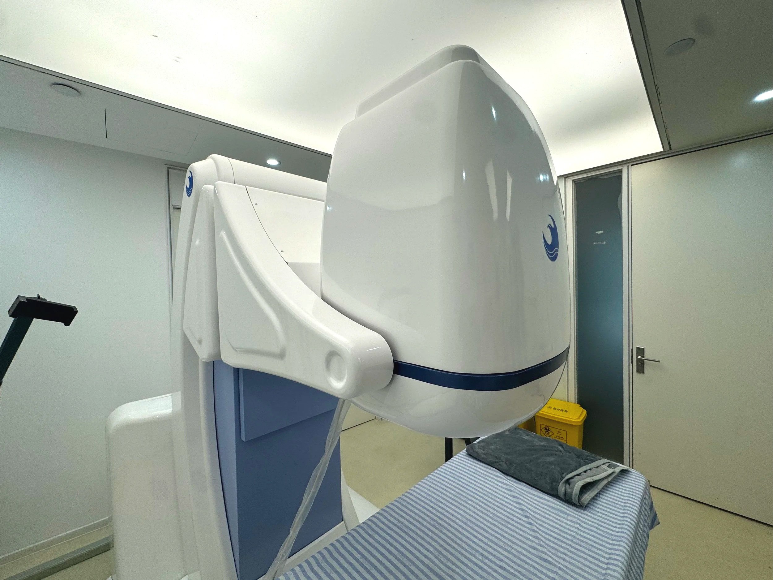
HY2900 Clinical Demonstration
Watch how HY2900 enables non-invasive, uterus-preserving treatment in a clinical setting.